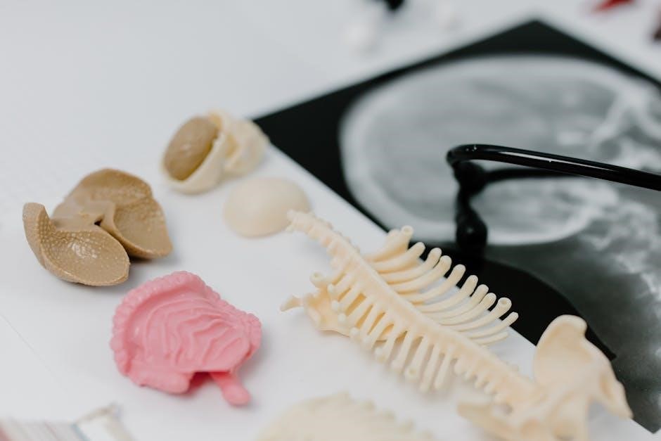
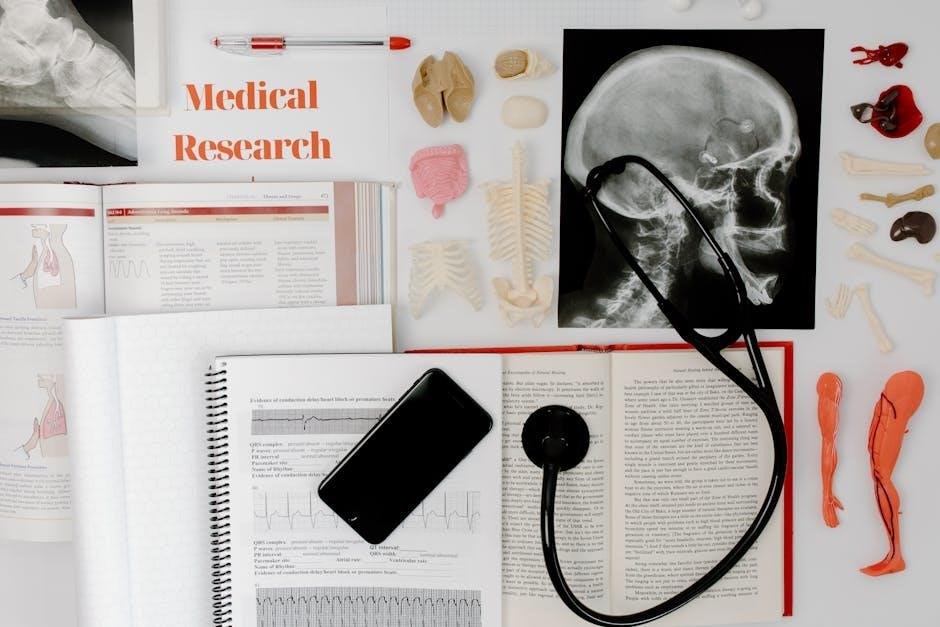
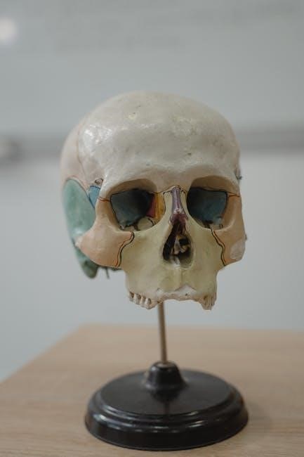
Anatomy and physiology coloring workbooks are interactive study tools that combine visual and tactile learning to master complex biological structures and processes. These workbooks offer a hands-on approach to understanding the human body, making learning engaging and effective. By coloring detailed illustrations, students can better retain information about the skeletal, muscular, and other key systems. This method enhances both knowledge retention and spatial awareness, providing a comprehensive foundation for anatomy and physiology studies. Ideal for students and professionals alike, these workbooks simplify complex concepts through creative, structured activities.
1.1 What is an Anatomy and Physiology Coloring Workbook?
An anatomy and physiology coloring workbook is an educational resource designed to help learners engage with complex biological concepts through creative, hands-on activities. It typically features detailed illustrations of the human body’s structures and systems, such as bones, muscles, organs, and tissues. By coloring these diagrams, users can visualize and internalize anatomical relationships, making abstract concepts more tangible. The workbooks often include labels, descriptions, and guided instructions to enhance understanding. This interactive approach fosters active learning, improving retention and spatial awareness. Unlike traditional textbooks, coloring workbooks provide a unique, artistic method for studying anatomy and physiology, catering to visual and kinesthetic learners.
1.2 Importance of Visual Learning in Anatomy and Physiology
Visual learning is crucial in anatomy and physiology, as these subjects rely heavily on understanding complex structures and their relationships. Diagrams, illustrations, and 3D models provide a clear framework for grasping spatial arrangements and functional connections. Visual aids help learners identify and differentiate between organs, tissues, and systems, enhancing comprehension and retention. Coloring workbooks leverage this by combining hands-on activity with visual engagement, making abstract concepts more tangible. This method is particularly effective for visual and kinesthetic learners, as it actively involves them in the learning process. By engaging multiple senses, visual learning fosters a deeper and more memorable understanding of anatomical and physiological principles.

Benefits of Using a Coloring Workbook for Anatomy and Physiology Study
Coloring workbooks actively engage students, enhancing focus and retention through visual encoding. They transform complex concepts into interactive, memorable experiences, fostering a deeper understanding of anatomical structures and physiological processes.
2.1 Engaging and Interactive Learning Experience
Anatomy and physiology coloring workbooks create an interactive and engaging learning environment. By coloring detailed illustrations, students actively participate in the learning process, making it more enjoyable and immersive. This hands-on approach allows learners to explore complex structures and systems, such as bones, muscles, and organs, in a visually stimulating way. The act of coloring helps students focus and retain information better, as it combines visual and tactile learning. This method is particularly effective for visual and kinesthetic learners, transforming abstract concepts into tangible experiences. It also fosters a sense of accomplishment as students see their work come to life, enhancing motivation and overall understanding.
2.2 Reinforcing Memory Through Color-Coding
Color-coding in anatomy and physiology workbooks is a powerful tool for reinforcing memory. By assigning specific colors to different structures or systems, learners can visually distinguish and remember complex details. This method leverages the brain’s ability to associate colors with information, making retention easier. For example, using red for arteries and blue for veins helps in quickly identifying cardiovascular structures. The psychological impact of color enhances recall, as vibrant visuals create lasting mental images. This technique not only aids in memorizing anatomical details but also helps in distinguishing between similar structures, making it an invaluable asset for effective learning and long-term retention of physiological concepts.
2.3 Developing Fine Motor Skills and Hand-Eye Coordination
Engaging with anatomy and physiology coloring workbooks helps develop fine motor skills and hand-eye coordination. The precise act of coloring intricate anatomical illustrations requires careful movements, enhancing dexterity and control. As users focus on staying within lines and shading specific areas, their ability to coordinate hand movements with visual perception improves. This skill is particularly beneficial for students and professionals in healthcare, where manual precision is essential. Regular practice with coloring tools strengthens muscle memory, making tasks like dissection or surgery more intuitive. Additionally, the repetitive motion of coloring can improve overall hand stability, further refining motor abilities. This makes coloring workbooks a practical tool for skill development beyond academic learning.

Key Anatomical Systems Covered in the Workbook
The workbook covers major anatomical systems, including skeletal, muscular, cardiovascular, respiratory, nervous, digestive, endocrine, urinary, and immune/lymphatic systems. Each system is detailed with precise illustrations for comprehensive understanding and retention.
3.1 Skeletal System: Bones and Joints
The skeletal system, comprising 206 bones, forms the structural framework of the body. Bones vary in shape, size, and function, from long bones like the femur to flat bones like the skull. Joints, where bones connect, allow for movement and stability. The workbook provides detailed illustrations of bone structures, such as the ribcage, vertebrae, and pelvis, as well as joint types like synovial and cartilaginous joints. Coloring these diagrams helps students differentiate between bone types and understand how joints facilitate movement. This section also highlights the role of ligaments and tendons in connecting bones, reinforcing the skeletal system’s dynamic and supportive role in the body.
3.2 Muscular System: Structure and Function
The muscular system consists of over 650 muscles, enabling movement, maintaining posture, and regulating body temperature. Muscles are categorized into three types: skeletal (voluntary), smooth (involuntary), and cardiac (heart-specific). Skeletal muscles attach to bones via tendons, facilitating voluntary movements, while smooth muscles line internal organs, controlling functions like digestion. Cardiac muscles are specialized for continuous, rhythmic contractions. The workbook features detailed illustrations of muscle groups, such as the biceps, quadriceps, and abdominal muscles, along with their attachments and functions. Coloring these diagrams helps students visualize how muscles interact with bones and other systems, reinforcing understanding of their roles in movement and overall bodily functions.
3.3 Cardiovascular System: Heart and Blood Vessels
The cardiovascular system, comprising the heart and blood vessels, is vital for circulating blood throughout the body. The heart, a muscular organ, pumps blood through arteries, veins, and capillaries, supplying oxygen and nutrients to tissues. Arteries carry oxygen-rich blood away from the heart, while veins return oxygen-poor blood. Capillaries facilitate nutrient and waste exchange. The workbook includes detailed illustrations of the heart’s chambers, valves, and vascular networks. Coloring these diagrams helps students visualize the system’s complexity, such as the distinction between pulmonary and systemic circulation. This interactive approach enhances understanding of blood flow and its essential role in maintaining life.
3.4 Respiratory System: Lungs and Airways
The respiratory system, essential for oxygen exchange, includes the lungs and airways that facilitate breathing. The trachea divides into bronchi, leading to bronchioles within the lungs, ending in alveoli where gas exchange occurs. Coloring detailed illustrations of the nasal cavity, pharynx, larynx, trachea, and lungs helps students visualize airflow and oxygen absorption. The workbook emphasizes the diaphragm’s role in expanding the chest cavity during inhalation. By coloring these structures, learners better understand how oxygen reaches the bloodstream and carbon dioxide is expelled. This interactive method reinforces the respiratory system’s critical function in maintaining life and overall bodily functions.
3.5 Nervous System: Brain, Spinal Cord, and Nerves
The nervous system is a complex network controlling bodily functions, comprising the brain, spinal cord, and nerves. The brain, the control center, consists of the cerebrum, cerebellum, and brainstem, regulating thought, movement, and involuntary actions. The spinal cord connects the brain to the body, transmitting signals through nerves. Coloring illustrations of neural pathways and structures helps learners visualize how electrical impulses travel, enabling functions like sensation, movement, and cognition. This interactive approach simplifies understanding of the nervous system’s intricate roles in communication and coordination, making it easier to grasp its essential functions in maintaining overall bodily harmony and responsiveness.
3.6 Digestive System: Organs and Processes
The digestive system is a complex process involving the breakdown and absorption of nutrients from food. Key organs include the mouth, esophagus, stomach, small intestine, and large intestine. The mouth initiates digestion with teeth and enzymes, while the stomach further breaks down food using acids and churning. The small intestine absorbs nutrients into the bloodstream, and the large intestine eliminates waste. Coloring diagrams of these organs and their functions helps learners visualize the digestive process, from ingestion to excretion. This interactive method enhances understanding of how enzymes, acids, and intestinal lining contribute to nutrient absorption, making the digestive system’s roles in sustenance and energy clear and memorable.
3.7 Endocrine System: Hormones and Glands
The endocrine system is a network of glands that produce and regulate hormones essential for various bodily functions. Key glands include the pituitary, thyroid, pancreas, adrenal glands, and reproductive organs like the ovaries and testes. Hormones, such as insulin, adrenaline, and estrogen, control processes like metabolism, energy levels, and growth. Coloring diagrams of these glands and their hormone pathways helps learners visualize how they interact to maintain homeostasis. This tactile approach enhances understanding of how the endocrine system influences growth, development, and overall health, making complex hormonal relationships more accessible and memorable for students studying anatomy and physiology.
3.8 Urinary System: Kidneys and Excretion
The urinary system, or renal system, is responsible for removing waste, excess fluids, and electrolytes from the body through urine production. The kidneys, primary organs of this system, filter blood to produce urine, which travels through the ureters to the bladder for storage and is expelled via the urethra. Coloring workbooks often highlight the kidneys’ structure, including nephrons, the functional units where filtration occurs. This visual learning tool helps students understand the process of urine formation, from glomerular filtration to tubular reabsorption. By coloring diagrams, learners can better grasp how the urinary system maintains homeostasis and overall bodily function through efficient waste removal and fluid balance regulation.
3.9 Immune/Lymphatic System: Defense Mechanisms
The immune and lymphatic systems work together to protect the body from pathogens, diseases, and foreign substances. The lymphatic system, comprising lymph nodes, spleen, and lymph vessels, filters fluids and aids in immune cell circulation. The immune system, including white blood cells like lymphocytes, identifies and neutralizes threats. Coloring workbooks often depict these systems’ interconnectedness, highlighting how lymphatic vessels transport immune cells and waste. By coloring diagrams of lymph nodes and spleen structures, learners can visualize how these systems detect and respond to antigens, reinforcing understanding of immune responses and disease prevention mechanisms. This interactive approach simplifies complex immune processes for easier retention and comprehension.
Physiological Processes Explained Through Coloring
Coloring workbooks simplify complex physiological processes like blood circulation, nerve impulses, and muscle function, transforming abstract concepts into visual, memorable experiences that enhance understanding and retention.
4.1 Cell Structure and Function
Coloring workbooks provide a visual approach to understanding cell structure and function, helping students identify and memorize components like the nucleus, mitochondria, and cell membrane. By coloring diagrams, learners can better grasp how cells operate, including processes like osmosis and protein synthesis. This method enhances spatial awareness and retention of complex cellular concepts. Interactive coloring also aids in understanding the roles of organelles and their interactions within the cell. Additionally, it helps students connect cellular functions to larger physiological processes, making it easier to build a foundation for advanced topics in anatomy and physiology. This engaging technique simplifies learning and reinforces key biological principles effectively.
4;2 Tissues and Their Classification
Tissues are groups of similar cells that perform specific functions, forming the building blocks of organs. Coloring workbooks simplify the classification of tissues into four primary types: epithelial, connective, muscle, and nervous. Epithelial tissues form linings and coverings, while connective tissues provide support and structure. Muscle tissues enable movement, and nervous tissues facilitate communication. Through detailed illustrations, students can color and differentiate these tissues, enhancing their understanding of their roles and locations within the body. This visual approach aids in recognizing tissue organization and function, making complex anatomical concepts more accessible and memorable for learners at all levels.
4.3 Body Fluids and Circulation
Body fluids, including blood, lymph, and interstitial fluid, play a crucial role in maintaining homeostasis. Coloring workbooks help visualize the circulatory system, highlighting how blood flows through arteries, veins, and capillaries. By coloring illustrations of the heart and blood vessels, students can better understand blood circulation and its role in delivering oxygen and nutrients. The lymphatic system, often colored differently, shows how it aids in returning fluids to the bloodstream. This interactive method enhances comprehension of fluid dynamics and circulation pathways, making complex physiological processes easier to grasp and remember for anatomy and physiology learners.
4.4 Nerve Impulses and Muscle Contractions
Nerve impulses and muscle contractions are fundamental to movement and communication within the body. Coloring workbooks provide detailed illustrations of neurons, synapses, and muscle fibers, allowing students to visualize how electrical signals propagate. By coloring neural pathways, learners can trace the flow of action potentials and understand synaptic transmission. Muscle contraction mechanisms, such as the sliding filament theory, are also illustrated, showing how actin and myosin fibers interact. This hands-on approach clarifies the connection between nerve signals and physical movement, making these complex processes more accessible and memorable for anatomy and physiology students.

Advanced Topics in Anatomy and Physiology
Explore complex systems like neuroanatomy, histology, and radiological imaging. Detailed sections on CT, MRI, and ultrasound diagnostics enhance understanding of advanced anatomical structures and physiological processes.
5.1 Regional Anatomy: Head, Neck, and Trunk
Regional anatomy focuses on the organized study of specific body regions, such as the head, neck, and trunk. The head includes structures like the skull, brain, and sensory organs, while the neck contains vital vessels and the thyroid gland. The trunk is divided into the thorax, abdomen, pelvis, and back, housing major organs like the heart, lungs, liver, and kidneys. Coloring workbooks often dedicate detailed sections to these regions, allowing learners to visualize and differentiate between layers of muscles, nerves, and blood vessels. This approach helps in understanding the spatial relationships and functional connections within these complex areas, enhancing anatomical comprehension and clinical application.
5.2 Cross-Sectional Anatomy: CT and MRI Imaging
Cross-sectional anatomy involves studying the body in slices, providing detailed views of internal structures. CT (Computed Tomography) and MRI (Magnetic Resonance Imaging) scans are essential tools for visualizing these sections. Coloring workbooks often include cross-sectional images, allowing learners to color-code tissues, organs, and abnormalities. This interactive approach helps users recognize anatomical landmarks and understand spatial relationships. By coloring CT and MRI images, students can differentiate between soft tissues, bones, and fluids, enhancing their ability to interpret diagnostic imaging. This method bridges anatomy with clinical practice, making it invaluable for medical professionals and students alike to grasp complex anatomical details in a visually engaging way.
5.3 Radiological Anatomy: X-Ray and Ultrasound
Radiological anatomy focuses on the study of internal structures using imaging techniques like X-rays and ultrasounds. X-rays are commonly used to examine bones, joints, and certain soft tissues, providing clear images of fractures or abnormalities. Ultrasound, on the other hand, utilizes sound waves to produce real-time images of organs and tissues, particularly useful for abdominal, pelvic, and fetal imaging. Coloring workbooks often incorporate these imaging modalities to help learners identify anatomical structures and pathological conditions. By coloring and labeling radiological images, students can enhance their understanding of diagnostic procedures and improve their ability to interpret clinical images, making it a valuable tool for both education and professional practice.

Tools and Resources for Effective Study
Anatomy and physiology coloring workbooks serve as essential tools, complementing 3D models, interactive apps, and online platforms to enhance visual and tactile learning experiences for students and professionals.
6.1 3D Anatomy Models for Visual Learning
3D anatomy models are powerful tools that complement coloring workbooks by providing an interactive, three-dimensional understanding of the human body. These models, often created by physicians and anatomy experts, allow users to explore structures like bones, muscles, and organs in detail. Platforms such as e-Anatomy and Anatomy Learning offer comprehensive libraries of 3D models, utilizing international anatomical terminology. Students can rotate, zoom, and dissect virtual structures, enhancing their spatial awareness and comprehension of complex anatomical relationships. These models are particularly beneficial for kinesthetic and visual learners, offering a dynamic way to study anatomy alongside traditional methods like coloring workbooks.
6.2 Interactive Anatomy Software and Apps
Interactive anatomy software and apps are cutting-edge tools that enhance learning by allowing users to explore the human body in immersive detail. Platforms like TeachMeAnatomy and e-Anatomy offer highly detailed, 3D models of anatomical structures, enabling users to zoom, rotate, and dissect virtual specimens. These tools often include features like labeling, quizzes, and virtual dissections, making them ideal for self-directed study. Many apps are accessible on both desktop and mobile devices, providing flexibility for learners. By integrating interactive features with traditional study methods, such as coloring workbooks, students can achieve a deeper understanding of anatomy and physiology. These tools are invaluable for visual and kinesthetic learners.
6.3 Online Anatomy Platforms and Communities
Online anatomy platforms and communities offer vast resources for anatomy and physiology learning, providing interactive tools, detailed guides, and opportunities for collaboration. Websites like TeachMeAnatomy and e-Anatomy host comprehensive libraries of anatomical images, 3D models, and study guides. These platforms cater to both students and professionals, offering quizzes, virtual dissections, and imaging resources like CT and MRI scans. Additionally, online forums and communities allow learners to discuss complex topics, share study tips, and collaborate on challenging concepts. By integrating these resources with a coloring workbook, students can enhance their understanding of anatomy and physiology through a combination of visual, tactile, and digital learning methods.

Tips for Using the Coloring Workbook Effectively
Consistently practice coloring to reinforce anatomical knowledge, use reference materials for accuracy, and balance coloring with active study techniques for optimal learning outcomes and retention.
7.1 Creating a Study Schedule
Creating a study schedule is essential for effective use of an anatomy and physiology coloring workbook. Begin by allocating specific times each day or week for coloring and review. Prioritize consistency to build a strong foundation of knowledge. Set realistic goals, such as completing one or two sections per session, and ensure each session includes both coloring and conceptual understanding. Use a planner or digital app to track progress and stay organized. Dedicate time for reviewing colored pages to reinforce memory and identify areas needing further study. Avoid cramming by spacing out sessions, and incorporate breaks to maintain focus and prevent burnout. Adjust the schedule as needed to accommodate learning pace and other commitments, ensuring it remains flexible yet structured. Regular review and adherence to the schedule will enhance learning outcomes and retention. Stay committed to maintain momentum and achieve long-term understanding of anatomy and physiology concepts through consistent practice.
7.2 Using Color-Coding Strategies
Using color-coding strategies in an anatomy and physiology coloring workbook enhances learning by organizing information visually. Assign specific colors to different structures, systems, or processes to differentiate them clearly. For example, use red for arteries and blue for veins to distinguish cardiovascular pathways. Consistency in color selection aids in recognizing patterns and relationships between components. Digital tools can also help maintain uniformity across pages. Color-coding reinforces memory by associating hues with specific functions or locations, making complex concepts easier to retain. Experiment with various palettes to create a visually appealing and educationally effective study resource. This method encourages active engagement and deeper understanding of anatomical details through vibrant, structured representation.
7.3 Reviewing and Revising Colored Pages
Regularly reviewing and revising colored pages in your anatomy and physiology workbook is crucial for reinforcing knowledge and identifying gaps. Set aside time to revisit completed sections, ensuring all details are accurate and well-understood. Use different colors to correct mistakes or add annotations, enhancing clarity and depth. This process helps solidify complex concepts and improves long-term retention. By revisiting your work, you can track progress, refine your understanding, and build confidence in your grasp of anatomical structures and physiological processes. Consistent review ensures that the information remains fresh and accessible, making it easier to apply in practical or exam settings.
Anatomy and physiology coloring workbooks provide a valuable, effective way to engage with complex topics, enhancing learning and retention through interactive, creative study. They serve as essential tools for understanding the human body, making them indispensable for students and professionals alike in mastering anatomy and physiology.
8.1 The Role of Coloring Workbooks in Modern Anatomy Education
Anatomy and physiology coloring workbooks play a pivotal role in modern education by providing an engaging, interactive method for mastering complex anatomical structures. These tools complement traditional textbooks and digital resources, offering a hands-on approach that enhances visual and tactile learning. Platforms like TeachMeAnatomy and Anatomy Learning integrate 3D models and interactive guides, while workbooks allow students to color and label structures, reinforcing spatial awareness and retention. This method is particularly effective for understanding regional anatomy and cross-sectional imaging, making it a valuable supplement to modern study tools and resources for both students and professionals in the field of anatomy and physiology.
8.2 Encouraging Lifelong Learning in Anatomy and Physiology
Anatomy and physiology coloring workbooks foster a culture of continuous learning by making complex concepts accessible and engaging. These tools cater to diverse learning styles, encouraging self-directed study and exploration. By integrating visual and tactile exercises, they help learners develop a deeper understanding of human anatomy, which is essential for both academic and professional growth. Platforms like TeachMeAnatomy and Anatomy Learning further support this journey by offering interactive resources and communities. Coloring workbooks, combined with digital tools, create a comprehensive learning ecosystem that inspires curiosity and motivates learners to explore anatomy and physiology throughout their lives, whether for personal interest or professional advancement.